

In the course of the XIV National Congress of the Spanish Skull Base Society.
- Its use as a tool for surgical planning allows a comprehensive approach to the most complex skull base pathologies.
- The combination of cutting-edge engineering with the most advanced medical techniques opens up a new range of possibilities in neurosurgery.
- The latest innovations in this field were presented at the National Congress of the Spanish Skull Base Society held in Alicante.
Madrid, October 30. Innovation and commitment to the development of state-of-the-art medical technology is one of the pillars underpinning advances in neurosurgery. Given the complexity of this specialty, it is essential to invest in new models and techniques that allow specialists to perform their work with increasing precision.
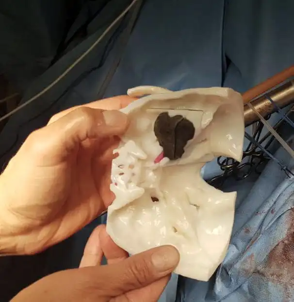
On the occasion of the celebration of the XIV National Congress of the Spanish Skull Base Society in Alicante, some of the most outstanding innovations in this field have been presented. The presentation on the latest advances in anatomical biomodels manufactured by 3D printing attracted great interest. This is a reliable, life-size reproduction of the case to be operated on. A real object that the surgeon can hold in his hand and observe from any angle.
A tool to increase surgical precision
Recent advances in biomedical engineering make it possible to generate 3D digital biomodels from 2D CT images using software. It is also possible to superimpose images obtained by MRI or other techniques on the CT images to obtain high-quality three-dimensional reconstructions. The process of creating each 3D biomodel is the result of hand-in-hand collaboration between the surgeon and the engineering team; it is not an automatic process, but the result of the application of a set of software tools under the auspices of the person who will subsequently perform the operation in the operating room.
“Having advanced 3D biomodels helps to plan the surgical intervention. Each case is unique and full of nuances, which is crucial because the procedure must be tailored to each individual patient. Individual and reliable knowledge of the anatomy and pathology allows the best access route to be chosen, difficulties to be anticipated and the case to be approached with more confidence. Collaboration with the Digital Anatomics team has allowed us to evolve in step with the latest technological advances in a fruitful way over the last few years,” explains Dr. David Santamarta, a neurosurgeon familiar with vascular and tumor skull base surgery, and a member of the Neurosurgery Service of the Complejo Asistencial Universitario de León.
“Throughout these last four years, only the collaboration with surgeons and the performance of countless tests have allowed us to reach the point where we are today, making the biomodels more and more useful and perfected. This process has led us, for example, to generate a complex biomodel of a Congenital Cholesteatoma, for the first time with multiple tissues and densities that has been really useful to achieve the success of the last intervention” concludes Alejandro Reyero, engineer in charge of leading the Digital Anatomics team.