PERSONALIZED SURGICAL GUIDES AND RADIATION-FREE PLANNING: DIGITAL ANATOMICS PARTICIPATES IN A SPINE SURGERY STUDY PUBLISHED IN THE EUROPEAN SPINE JOURNAL
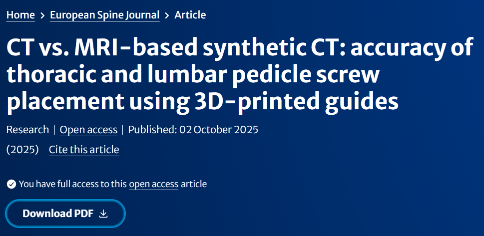
A study recently published in the European Spine Journal evaluated the accuracy of pedicle screw placement in the thoracic and lumbar spine using 3D-printed surgical guides based on either conventional CT images or synthetic CT (sCT) derived from MRI. The study aimed on reducing radiation exposure in surgical planning.
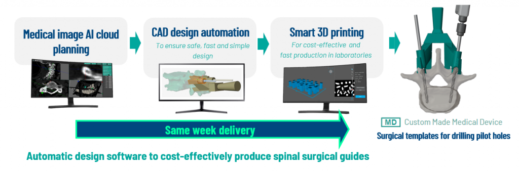
Digital Anatomics technology was used in all key phases of the study, including:
- Automatic vertebral segmentation and pedicle screw trajectory planning using our AI-based software.
- Automated CAD design generation of the surgical guides.
- Manufacturing and supply of the guides and 1:1 anatomical models used during the procedures.
The study was conducted on cadaveric specimens and compared two groups of guides, one based on CT and one on sCT imaging. A total of 62 K-wires were inserted across 31 vertebrae, and deviations from the preoperative planning were measured in both angular and linear terms.
No statistically significant differences were observed between the two imaging methods.
The median translational deviation at the screw entry point was below 1.1 mm, and below 1.2 mm at the pedicle level, with median angular deviations under 2.5°.
These values confirm the high precision of patient-specific 3D-printed guides, even when based on MRI-derived synthetic CT.
According to published data, computer-assisted navigation and robotic systems typically report entry-point and pedicle deviations in the range of 1–2 mm and angular deviations of 2–6°, placing the present results well within the lower range of currently reported accuracies.
The rate of pedicle wall breach was also evaluated, with no significant deviations from the plan detected in either group.
This research validates the feasibility of radiation-free planning based on MRI-derived images, and demonstrates that a fully digital workflow, from automatic segmentation to physical guide production, can be executed with high precision.
For Digital Anatomics, this collaboration represents a technical and scientific validation of our tools, showing their applicability with both conventional imaging and new modalities such as sCT. It also reinforces the value of personalized surgical solutions in spine surgery, providing support to surgeons without replacing their clinical judgment.