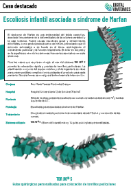
Dra. María Teresa Menéndez Crespo
Hospital Universitario 12 de Octubre (Madrid, Spain)
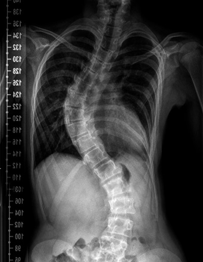
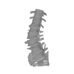
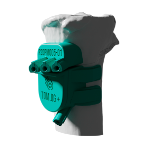
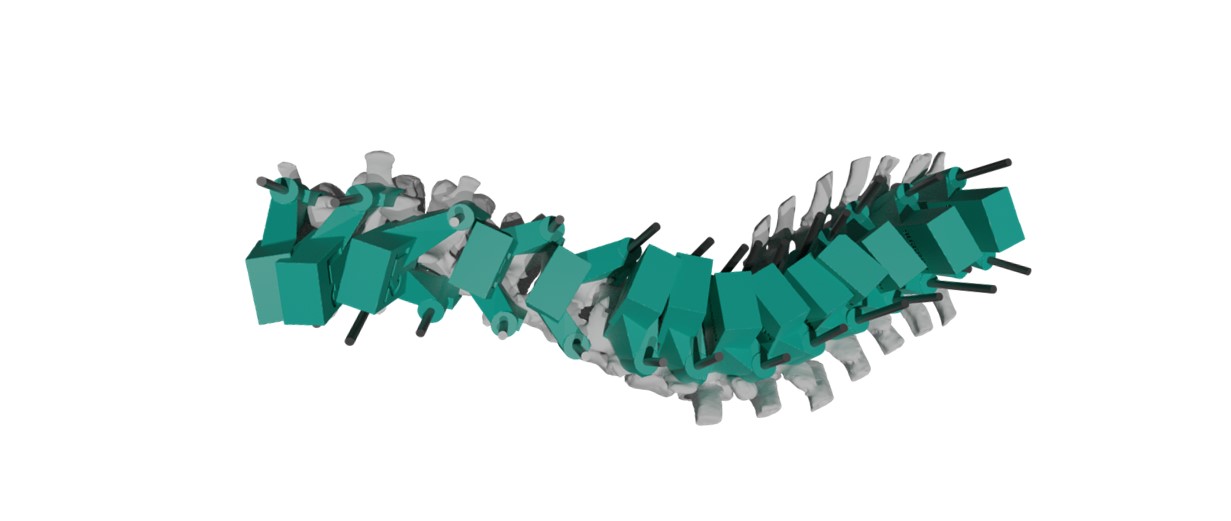
14-year-old girl; she presents scoliosis with right thoracic curvature of 70˚ and left lumbar of 43˚.
Severe scoliosis associated with Marfan syndrome
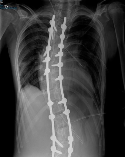
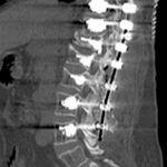
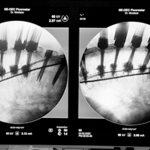
Instrumented posterior arthrodesis surgery from T3 to L4 and correction of the curves.
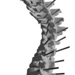
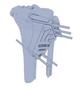
TOR JIG ® S : Anatomic biomodel and 14 personalized surgical guides